Coronary Angiography & Angioplasty
- This procedure is meant for the diagnosis and treatment of ischemic heart disease.
- Minimal invasive procedure.
- Local anesthetics is administered to the site of catheter entry.
- Catheter is inserted through the femoral or radial artery.
- Radiopaque dye is injected into the coronary arteries via the wire in order to get a visualization of the vessels with the help of a fluoroscope machine.
- The doctor can make a diagnostic decision about whether there are significant lesions in the coronary arteries or not.
- Guide wire is sent through the catheter to the heart vessels.
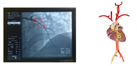
- In case of significant lesion, the doctor will decide to perform balloon/stent angioplasty.
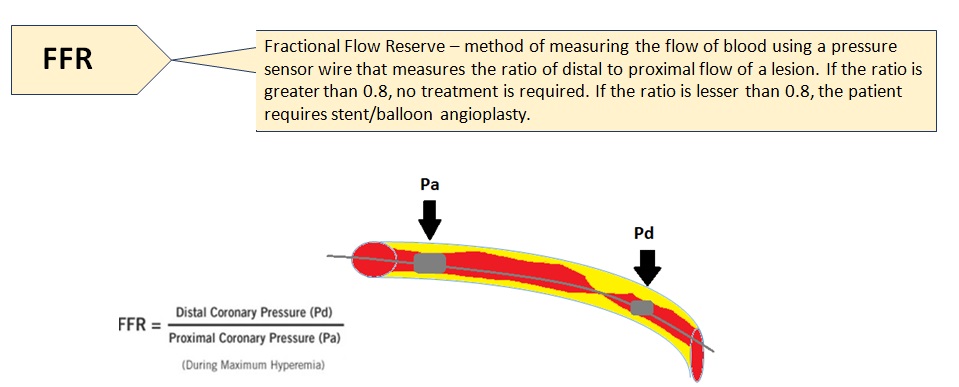
- The doctor can also use an FFR catheter in order to get more information about the flow of blood proximal to the lesion relative to distal to the lesion, especially when the lesion does not seem significant on the X-ray images or are borderline.
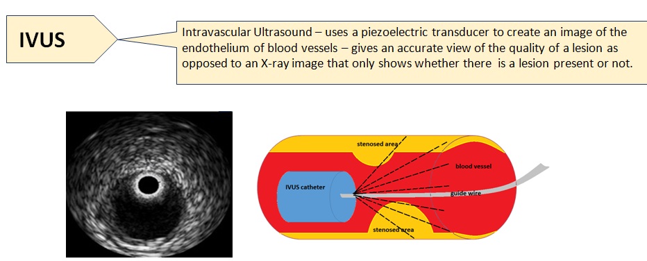
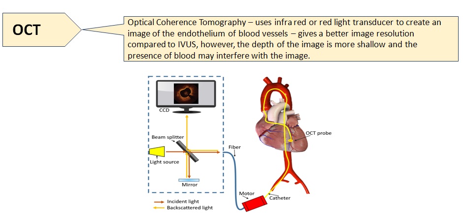
- In case of suspected calcification lesions, or borderline lesions IVUS or OCT catheters may be used.
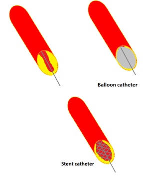
- The hollow balloon catheter can be inserted into the back of the coronary guidewire – the balloon catheter is on the inside of the balloon catheter and slides forwards.
- The balloon is navigated to the site of stenosis along the coronary guidewire and the inflated once it reached there.
- The balloon causes the artery wall to stretch as it inflates, and opens up and compresses the plaque.
- If there was a stent attached to the balloon, it will latch onto the stretched artery wall and hold it in place – implanted.
