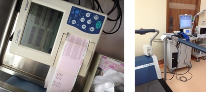
ECG
Electrocardiographs
- An electrocardiograph is a machine that is used to perform electrocardiography (process of recording), and produces the electrocardiogram(ECG or EKG).
- Because of low ECG voltages that is measure across the body (100mv to 1mv) it necessitates a low noise circuit and instrumentation amplifiers are key.
- Before, electrocardiographs were constructed with analog electronics but Today, they use analog-to-digital converters to convert to a digital signal that can then be manipulated with digital electronics.
- There are some other components to the electrocardiograph:
- Safety features that include voltage protection for the patient and operator. (the heart is sensitive to the AC frequencies typically used for mains power (50 or 60 Hz))
- Defibrillation protection.
- Electrostatic discharge is similar to defibrillation discharge and requires voltage protection up to 18,000 volts.
- The right leg driver circuitry can be used to reduce common-mode interference (typically the 50/60 Hz mains power).
- Typical design for a portable electrocardiograph is a combined unit that includes a screen, keyboard, and printer on a small wheeled cart. The unit connects to a long cable that branches to each lead which attaches to a conductive pad on the patient.
- Lastly, the electrocardiograph may include a rhythm analysis algorithm that produces a computerized interpretation of the electrocardiogram. Included in this analysis is computation of common parameters that include PR interval, QT duration, corrected QT (QTc) duration, PR axis, QRS axis, and more.
- Earlier designs recorded each lead sequentially but current designs employ circuits that can record all leads simultaneously.
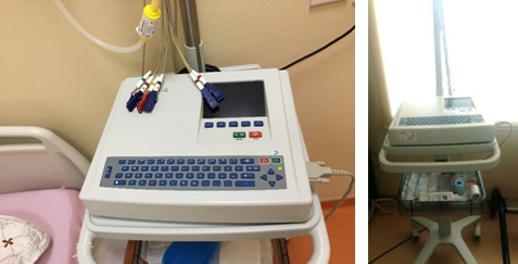
History
1903
Willem Einthoven
A Dutch doctor and physiologist, invented the first practical electrocardiogram and received the Nobel Prize in Medicine in 1924 for it
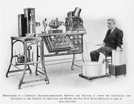

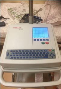
NOW
Modern ECG machine
has evolved into compact electronic systems that often include computerized interpretation of the electrocardiogram.
An ECG can help detect:
- Arrhythmias – where the heart beats too slowly, too quickly, or irregularly
- Coronary heart disease – where the hearts blood supply is blocked or interrupted by a build-up of fatty substances
- Heart attacks – where the supply of blood to the heart is suddenly blocked
- Cardiomyopathy – where the heart walls become thickened or enlarged
A series of ECGs can also be taken over time to monitor a person already diagnosed with a heart condition or taking medication known to potentially affect the heart.
Understanding ECG Waveform
 F P T D.jpg)
The ECG Paper
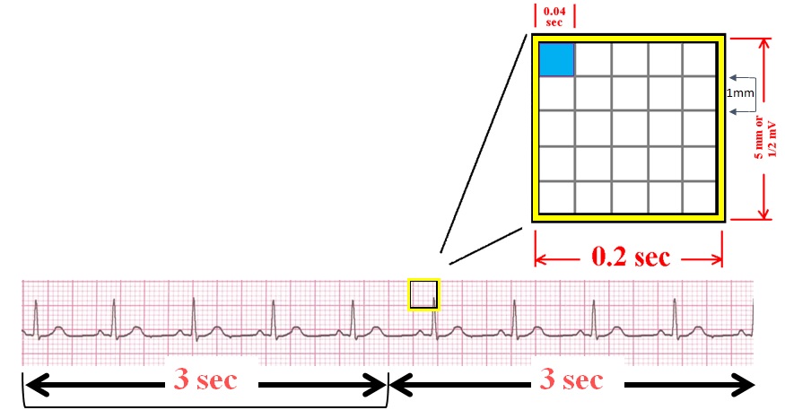
The graph paper that the ECG records on is standardized to run at 25mm/second, and is marked at 1 second intervals on the top and bottom.
- Horizontally : correlates the length of each electrical event with its duration in time
- One small box (Blue) - 0.04 s
- One large box (yellow) - 0.20 s
- Vertically
- One large box (yellow) - 0.5 mV
- Every 3 seconds (15 large boxes) is marked by a vertical line.
- This helps when calculating the heart rate.
- Duration of a waveform, segment, or interval is determined by counting the blocks from the beginning to the end of the wave, segment, or interval.
Electrodes
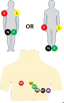
Usually consist of a conducting gel, embedded in the middle of a self-adhesive pad onto which cables dip. Ten electrodes are used for 12-lead ECG.
The limb electrodes
RA - On the right arm, avoiding thick muscle
LA - On the left arm this time.
RL - On the right leg, lateral calf muscle
LL - On the left leg this time.
The 6 chest electrodes
V1: Fourth intercostal space to the right of the sternum.
V2: Fourth intercostal space to the Left of the sternum.
V3: Directly between leads V2 and V4.
V4: Fifth intercostal space at midclavicular line.
V5: Level with V4 at left anterior axillary line.
V6: Level with V5 at left midaxillary line. (Directly under the midpoint of the armpit)
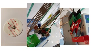
Electrodes…
- Two common electrodes used are :
- Flat paper-thin sticker
- Self-adhesive circular pad
- The former are typically used in a single ECG recording while the latter are for continuous recordings as they stick longer.
- Each electrode consists of an electrically conductive electrolyte gel and a silver/silver chloride conductor. The gel typically contains potassium chloride — sometimes silver chloride as well — to permit electron conduction from the skin to the wire and to the electrocardiogram
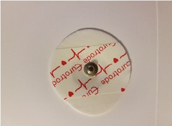
leads
- A "lead" is not the same as an "electrode".
- Whereas an electrode is a conductive pad in contact with the body that makes an electrical circuit with the electrocardiograph, a lead is a connector to an electrode. Since leads can share the same electrode, a standard 12-lead EKG happens to need only 10 electrodes.
- A lead is slightly more abstract and is the source of measurement of a vector.
- In medical settings, the term leads is also sometimes used to refer to the electrodes themselves, although this is not technically a correct usage of the term, which complicates the understanding of difference between the two.
The 12-Leads
- The 12-Lead ECG sees the heart from 12 different views.
- Therefore, the 12-Lead ECG helps you see what is happening in different portions of the heart.
- The rhythm strip is only 1 of these 12 views.
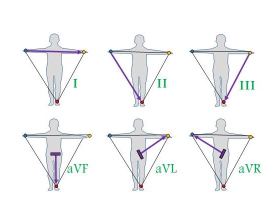
The 12-leads include :
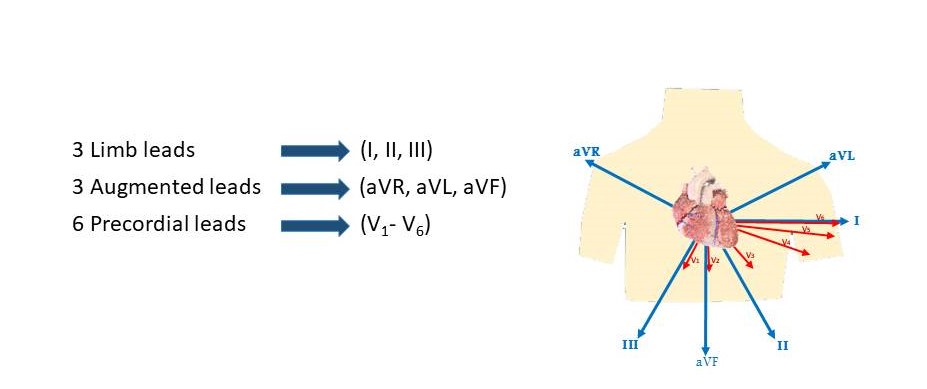
Contiguity of leads

aVR => Right atrium
Types of ECG
There are three main types of ECG:
- A resting ECG – carried out while patient is lying down in a comfortable position
- A stress or exercise ECG – carried out while patient is using an exercise bike or treadmill
- An ambulatory ECG – the electrodes are connected to a small portable machine worn at your waist so your heart can be monitored at home for one or more days
- The type of ECG recommended for patient will depend on the symptoms and the heart problem suspected.
- For example, an exercise ECG may be recommended if your symptoms are triggered by physical activity, whereas an ambulatory ECG may be more suitable if your symptoms are unpredictable and occur in random, short episodes.
