Intravascular Ultrasound (IVUS)
What is IVUS?
- Intravascular ultrasound (IVUS) is a catheter-based imaging technique providing real-time high resolution images of the vessel wall and lumen.
- Current IVUS catheters have a center frequency of 20-40 MHz.
- Sound waves bounce back with varying intensity and different time delays according to the density of the plaques they have encountered.
- Axial resolution is approximately 150 μm and the lateral resolution 300 μm.
- Grayscale IVUS allows robust quantitative measurements including lumen, vessel, and plaque area.
- PCI with the guidance of grayscale IVUS has been demonstrated to reduce incidence of restenosis, stent thrombosis and cardiovascular events after PCI compared to angiographic guidance.
- Recently, IVUS has been increasingly used as the gold standard to evaluate natural history of coronary atherosclerosis under medical therapies.

- The beam remains fairly parallel for a distance (near field) and then begins to diverge (far field)
- The quality of ultrasound images is better in the near field because the beam is more parallel and the resolution greater
- Therefore, larger transducers with lower frequencies are used for examination of large vessels because they create a deeper near field
IVUS Pullback System
Motorized Transducer Pullback
- Advantages
- Steady catheter withdrawal
- Creates length and volumetric measurements
- Uniform images
- Reproducible images
- Disadvantages
- May provide inadequate examination in regions of interest due top-reset speed
Manual Transducer Pullback
- Advantages
- Ability to concentrate on a specific region of interest
- Disadvantages
- Possibility of skipping over significant pathology by irregular pullback
- Potential inaccuracies in length and volume measurements
- The time delay of reflected ultrasound waves is translated into spatial image information while, the intensity is converted to an intensity map encoded by a grayscale.
- Dense targets such as calcified plaques are bright white in the IVUS image and the least dense plaques such as the medial layer in the vessel appear black.
- IVUS allows for precise measurement of the plaque burden by visualizing plaque topography. Also, it shows the shape of the lumen as well as the thickness of layers of the wall. Dense elements are shown as brighter white while the less dense elements are shown as darker.

IVUS Systems
The IVUS equipment consists of:
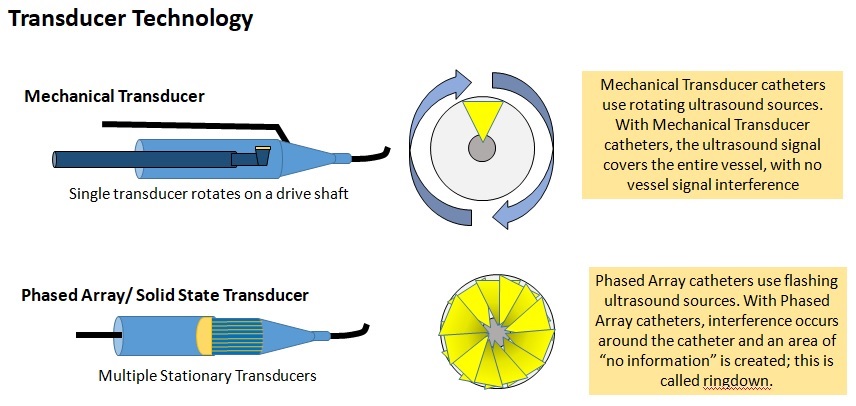
- Catheter (incorporating a miniaturized transducer)
- Console (reconstruct and display the image)
IVUS Integrated System
- Intended Advantages
- Immediate access
- Tableside control
- Save procedure room space
- May serve as a platform for other/new technology
- Disadvantages
- Not portable and Installation required
IVUS Cart System
- Intended Advantages
- Mobility
- No installation requirements
- Table side controller
- Disadvantages
- Transporting between rooms
- Space and availability
- Start up time
Limitations of grayscale IVUS
- Limited axial resolution
- Invasive procedure
- Currently limited to the assessment of patients who require CAG for a clinical indication
- Conventional IVUS provides a suboptimal characterization of the composition of atherosclerotic plaque (Inaccuracy in the assessment of plaque composition)