Optical Coherence Tomography (OCT)
Introduction
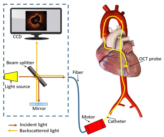
- Intravascular OCT technique measures the intensity of the back-reflected light.
- To minimize the light energy absorption by vessel wall components, 1300 nm wavelength (near-infrared light, 1250 to 1350 nm) is used.
- OCT can provide images with ultra-high resolution.
- Compared to IVUS, OCT provides images of in vivo plaques with approximately 10 times higher resolution which is up to 10 μm in an axial resolution and to 20 μm in a lateral resolution. Since OCT has shallow penetration depth of imaging, this modality is superior to monitor plaque microstructures below the endothelial surface.
Four major cardiovascular applications for OCT systems are:
1. Stent visualization
2. Vulnerable plaque assessment
3. Drug and device development
4. Peripheral artery disease visualization
How it works?
- It measures the intensity of reflected light and compares it with a reference. The reference is obtained by a mirror reflection on an arm.
- The mirror is dynamically translated in order to achieve cross correlation at incremental penetration depths in the tissue.
- High resolution images ranging from 4 micron to 20 micron can be achieved with a penetration depth of up to 2 mm.
- The frame rate is about 15 frames/sec.
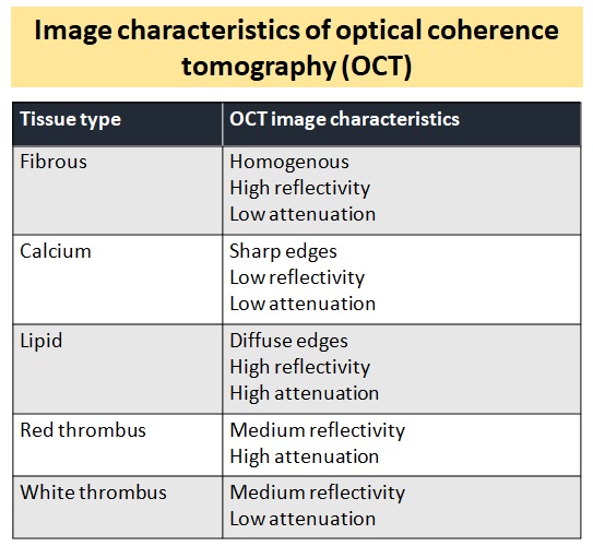
- Lipid pools generate decreased signal intensity compared to fibrous regions. Compared to IVUS, OCT demonstrates superior delineation of the thin caps or tissue proliferation.
There is two type of OCT sytems:
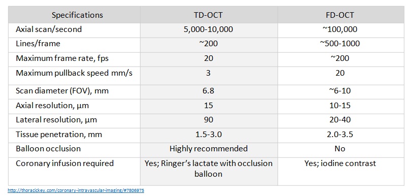
Time-domain OCT system (TD-OCT) : first generation (2006)
Fourier-domain OCT (FD-OCT) : new generation (2008)

Comparison of Time-Domain Optical Coherence Tomography and
Fourier-Domain Optical Coherence Tomography

OCT image acquisition
OCT cannot image through a blood field, and it requires clearing or displacing blood from the lumen by flushing.
There are two basic techniques:
- The occlusive technique.
- During image acquisition, coronary blood flow is stopped by inflating a proximal occlusion balloon and flushing the crystalloid solution at the rate of 0.5–1.0 ml/sec down the coronary artery.
- Non-occlusive technique.
- This is recently developed and does not need proximal balloon occlusion. During image acquisition the image wire pull back is performed at fast speed with simultaneous injection of contrast through the guiding catheter.

Limitations of OCT
- Poor tissue penetration(depth of 2 to 3 mm) and interference from blood.
- Due to this limited penetration, it is difficult to evaluate the entire amount of plaques (beyond the internal elastic lamina).
- This imaging is not suitable for visualizing atherosclerotic plaques in large arteries.
- Infusion of contrast medium is required during OCT imaging because infrared light does not penetrate red blood cells. This feature makes it difficult to use this imaging in patients with kidney disease.