Pulse Oximetry (SPO2)
Introduction
- Pulse oximetry is the continuous non‐invasive measurement of arterial haemoglobin oxygen saturation usually using a finger.
- The measurement is based on the Beer‐Lambert law: optical absorbency is proportional to the thickness of the medium and the concentration of the substance being measured.
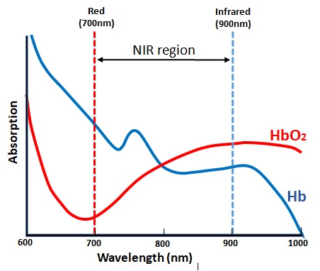
- Both red and infrared light are transmitted through the tissue with pulsatile blood flow. Oxygenated and deoxygenated haemoglobin differ in their capacity to absorb red (600–750 nm wavelengths) and infrared light (850–1000 nm wavelengths): oxygenated haemoglobin absorbs more infrared light than deoxygenated haemoglobin which, in turn, absorbs more red light than oxygenated haemoglobin.
- Comparison of absorbance at two different wavelengths (for example, 660 and 940 nm) enables estimation of the relative concentrations of oxygenated and deoxygenated haemoglobin.
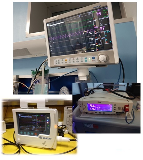
Monitor
- The monitor contains the microprocessor and display. The display shows the oxygen saturation, the pulse rate and the waveform detected by the sensor.
- The monitor is connected to the patient via the probe. During use, the monitor updates its calculations regularly to give an immediate reading of oxygen saturation and pulse rate.
- The audible beep changes pitch with the value of oxygen saturation and is an important safety feature.
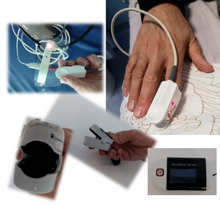
Probe
- The oximeter probe consists of two parts, the light emitting diodes (LEDs) and a light detector (called a photo-detector). Beams of light are shone through the tissues from one side of the probe to the other.
- The blood and tissues absorb some of the light emitted by the probe. The light absorbed by the blood varies with the oxygen saturation of haemoglobin. The photo-detector detects the light transmitted as the blood pulses through the tissues and the calculates a value for the oxygen saturation (SpO2).
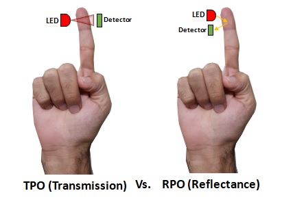
Pulse Oximetry can be done using two methods
- Reflectance Oximetry
- Transmittance Oximetry
- In case of reflectance oximetry, the two LEDs and the photodiode are on the same side. Here, the light moves through the skin, muscle and blood vessel, and is reflected back from the bone. Reflectance oximetry has low signal to noise ratio and difficult to set up.
- In case of transmittance oximetry, the two LEDs and the photodiode are on the opposite side of the finger. Here, the transmitted light is detected by the photo diode, and is found to have higher signal to noise ratio.
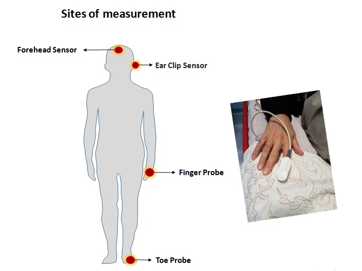
Limitation of pulse oximetres
- Pulse oximeters cannot distinguish between carboxyhaemoglobin or methaemoglobin and oxyhaemoglobin because their absorption spectra are similar.
- Occasionally, dark fingernail polish or very dark skin will cause the oximeter to under‐read by up to 5%.
- The ECG monitored heart rate should match that of the pulse oximeter; a difference in the readings implies that the pulse oximeter is not detecting arterial pulsation or that an artefact is contaminating the signal.
- Cardiac arrhythmias do not usually affect the accuracy of pulse oximetry but the use of vasoactive drugs may lead to under‐reading of the SpO2.