Treatment of congenital defects
Septal occluder device for covering septal holes
- Area of access is cleaned and/or shaven.
- Atrial septal defect – incision to the right internal jugular vein to access the pulmonary artery.
- Ventricular septal defect – incision in the femoral artery to access the aorta.
- A needle with syringe is inserted into the artery/vein.
- The syringe is removed (leaving the needle still in the blood vessel) and a short guidewire is inserted into the blood vessel via the needle.
- Once the short guidewire is in, the introducer sheath is inserted into the vessel along the guidewire.
- Once the introducer sheath is secure in place, the guidewire is removed and the guide catheter (J wire) is inserted through the introducer sheath.
- The guide catheter is navigated towards the heart, then the aortic arch/pulmonary artery using C-arm x-ray machine that provides real-time fluoroscopic images.
- Radiopaque contrast media is injected through a port in the introducer sheath into the guide catheter to provide a view of the heart vessels.
- Heparin is injected into the artery to prevent thrombosis or clotting.
- The wire that contains the septal occluder device is inserted into the blood vessel through the guide catheter, and then navigated to the hole in the atria/ventricles
- The device the passed through the septal hole and the disk on the end of the wire is opened and secured on one side of the hole.
- Then the disk on the other side of the defect is also opened and secured into place – the hole is covered on both sides now.
- Once the disks are implanted properly, the cable that is attached to the septal occlude device is detached and removed.
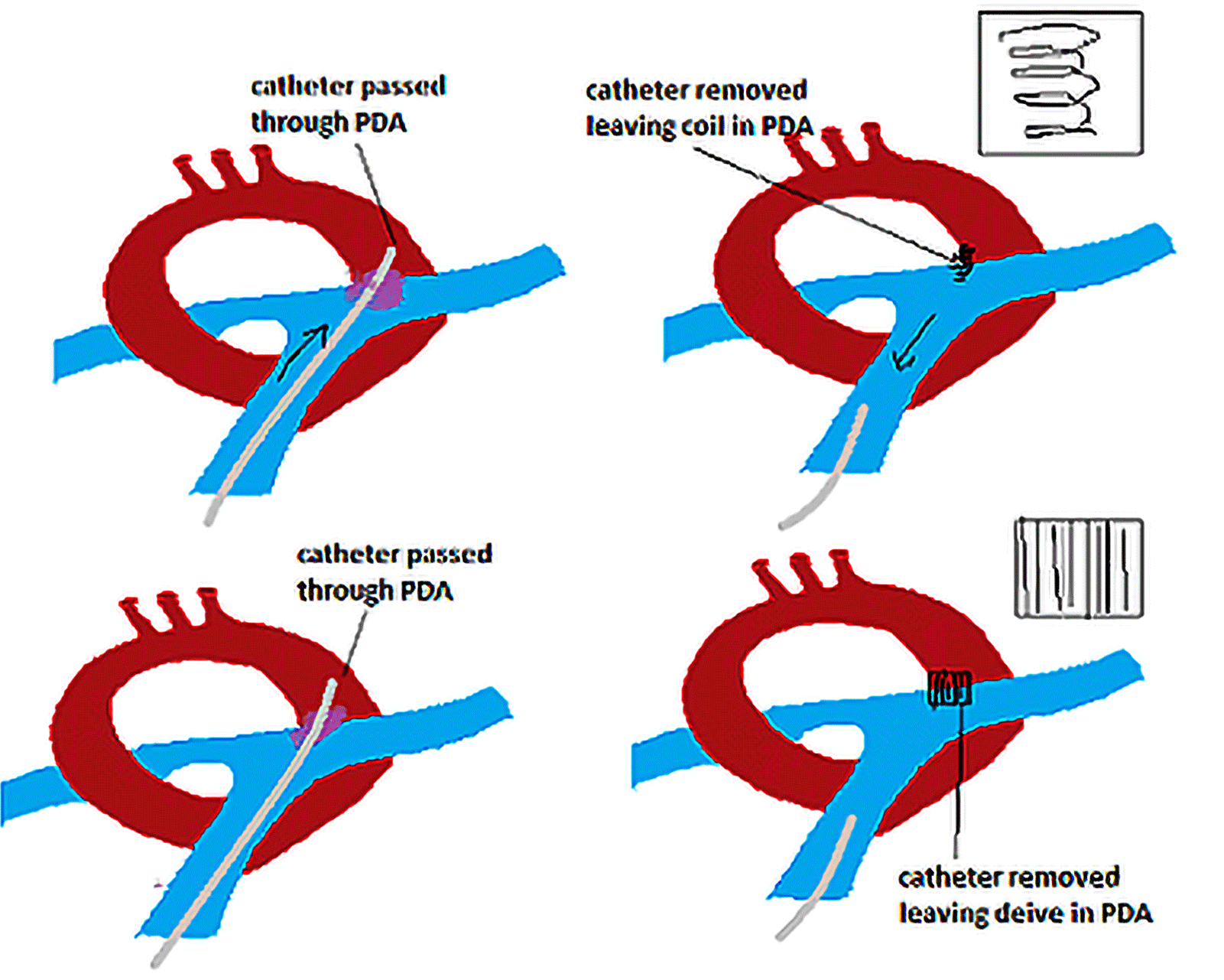
Patent ductus arteriosus
- Area of access is cleaned and/or shaven – femoral artery for access to the descending aorta.
- A needle with syringe is inserted into the artery.
- The syringe is removed (leaving the needle still in the blood vessel) and a short guidewire is inserted into the blood vessel via the needle.
- Once the short guidewire is in, the introducer sheath is inserted into the vessel along the guidewire.
- Once the introducer sheath is secure in place, the guidewire is removed and the guide catheter (J wire) is inserted through the introducer sheath.
- The guide catheter is navigated towards the heart, then the aortic arch/pulmonary artery using C-arm x-ray machine that provides real-time fluoroscopic images.
- Radiopaque contrast media is injected through a port in the introducer sheath into the guide catheter to provide a view of the heart vessels.
- Heparin is injected into the artery to prevent thrombosis or clotting.
- The wire that contains the septal occluder device is inserted into the blood vessel through the guide catheter, and then navigated to the hole in the aortic arch.
- The device the passed through the septal hole and the disk on the end of the wire is opened and secured on one side of the hole between the aorta and the pulmonary artery.
- Once the disks are implanted properly, the cable that is attached to the occluder device (occluder delivery system) is detached and removed.